CASE
HISTORY / CLINIC:
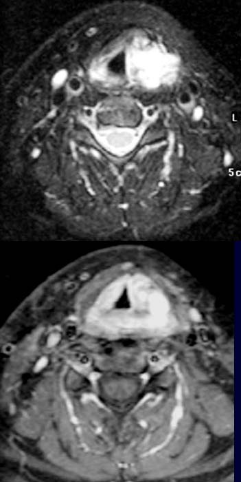
32-year-old patient with hoarseness and dysphagia.
FINDINGS:
The
T2-weighted fat
suppression image (above)
depicts a mass of
high signal intensity around the larynx and a mass of lesser signal intensity
on the right side of the larynx. Compression of the larynx lumen from the left
and slight displacement of the larynx to the right. The mass is relatively well
circumscribed and doesn't extend to the sheath
of vessels and nerves. The post-contrast T1-weighted fat
suppression image
(below) demonstrates an intensely enhancing, well circumscribed mass encircling
the larynx and growing mainly on the left side. Visualization of some central reduction
of signal intentensity.
DIAGNOSIS:
Larynx hemangioma
DIFFERENTIAL DIAGNOSIS:
Hemangiosarcoma
Web
resources:
1) Amersham Health
2) Medecoinfo