CASE
HISTORY / CLINIC:
24-year-old female with dysphagia.
FINDINGS:
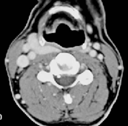
The CT image at the level of the hyoid demonstrates a strongly enhancing mass between
the greater horn of the hyoid and
the sheath
of vessels and nerves. This constricts the hypopharynx dorsally on the right.
The mass appears homogeneouly hyperdense.
DIAGNOSIS:
Ectopic right lobe of the thyroid gland lying extremely cranial.
DIFFERENTIAL DIAGNOSIS:
Other heavily vascularized benign masses.