CASE
HISTORY / CLINIC:
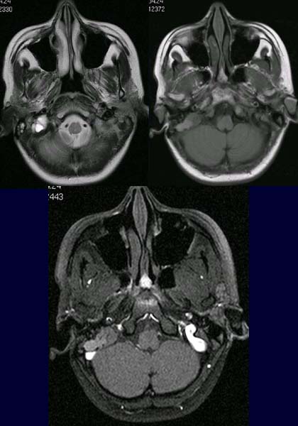
47-year-old patient with pronounced right-sided hearing impairment. No otorrhea.
FINDINGS:
The
T2-weighted MRT image (above right) demonstrates a lobed mass
lesion of mainly high signal intensity around the petrous pyramid.
Mass
lesion also shows a relatively high signal intensity in the non-contrast
T1-weighted image (above right).
In the post-contrast T1-weighted image after fat suppression (below) the mass
lesion basically appears unchanged in comparison to the non-enhanced image,
such that there is no evidence of contrast absorption.
DIAGNOSIS:
Cholesterol
granuloma
DIFFERENTIAL
DIAGNOSIS:
Cholesteatoma
Web
resources: