CASE
HISTORY / CLINIC:
Patient with paralysis of the right facial nerve for some months now. Buccal,
forehead, and eye branches are affected.
A small whitish swelling can be seen behind the intact eardrum in the upper posterior
portion. Normal findings of ear microscopy.
FINDINGS:
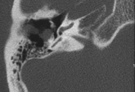
The non-contrast CT image through the petrous bone demonstrates a mass
lesion along the portion of the facial nerve that lies in the middle ear.
The ossicular
chain is not affected.
DIAGNOSIS:
Neurofibroma of the facial nerve.
DIFFERENTIAL DIAGNOSIS:
Neurinoma of the facial nerve
Web resources::