CASE
HISTORY / CLINIC:
Patient with recurring left-sided otorrhea as well as significant left-sided hearing
impairment/hearing loss.
FINDINGS:
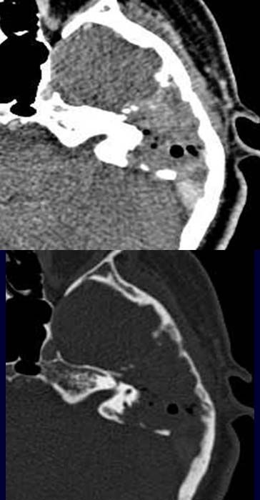
Demonstration of a hyperdense
mass lesion in the post-contrast axial CT
image through the petrous bone at soft tissue window (above). Mass lesion has
eroded structures in the middle ear and partly inner ear structures. No pneumatization
of the middle ear and petrous air cells as well as portions of the mastoid. The
bony erosion in
the middle and posterior cranial fossa as well as the invasion
of the inner ear are even better demonstrated in the bony window (below).
DIAGNOSIS:
Eosinophilic granuloma
DIFFERENTIAL DIAGNOSIS:
Malignant otitis externa.
Malignant tumor.
Web resources: