CASE
HISTORY / CLINIC:
55-year-old patient with recurring acute hearing loss on the left, the last episodes
of which occured last year and 8 weeks ago. A steep drop of the upper tones on
the left of the audiogramm
Normal fingings of ear microscopy. Failure of the equilibrium organ in the equilibrium
test.
FINDINGS:
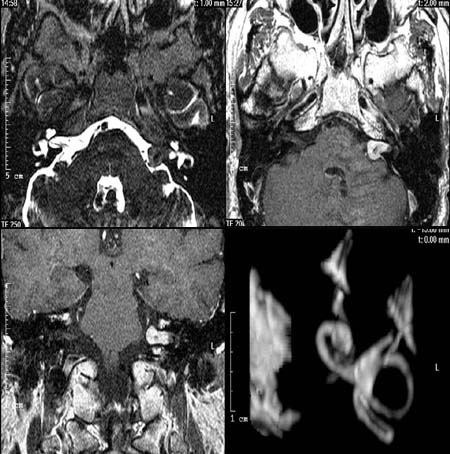
The CISS-Sequence (top left) demonstrates a displacement of the cerebrospinal
fluid around the left cerebellopontine angle and the left internal auditory meatus
depicting a hypodense
mass lesion. The mass lesion in the cerebellopontine angle and internal auditory
meatus shows a significant
signal enhancement in the post-contrast T1-weighted axial image (top right).
This hyperdense
mass lesion in the left cerebellopontine angle and internal auditory meatus
is also visualized in the post contrast T1-weighted coronary image (below left).
Excellent demonstration of the endolymph-filled semicircular
canals and the cochlea with
it's basal helices in the 3-D image (below right). No visualization of loss
in signal intensity in the fluid-filled cerebellopontine angle and internal
auditory meatus.
DIAGNOSIS:
Left-sided acoustic neurinoma
DIFFERENTIAL DIAGNOSIS:
Meningeoma
Web resources:
1) MedizInfo
2) Medicine worldwide
3) Netdoctor
4) Dr. Roepert
5) Onkologie.de
6) Amersham Health
7) Chorus
8) MRX