CASE
HISTORY / CLINIC:
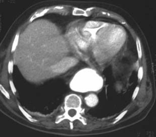
61-year-old patient with mild retrosternal pain.
FINDINGS:
CT scan of the abdomen after oral and i.v. contrast application:
A well defined plumply
contrast-filled retrocardial structure that
is also evident on the ventral side of the thoracic
aorta is visualized with patient lying supine. Homogeneous dorsobasal hyperintense
shadow on the right with discrete contrast
enhancement of the surrounding lung parenchyma.
DIAGNOSIS:
Small dorsobasal pleural
effusion on the right with surrounding dystelectasis. Axial
hiatal hernia.
Web
resources:
1) Jend
2) Endoskopischer Atlas
3) Learning radiology
4) Huber
5) RWTH Aachen
6) Medicine worldwide
7) AWMF
8) Chorus
9) AHC-Consilium