CASE
HISTORY / CLINIC:
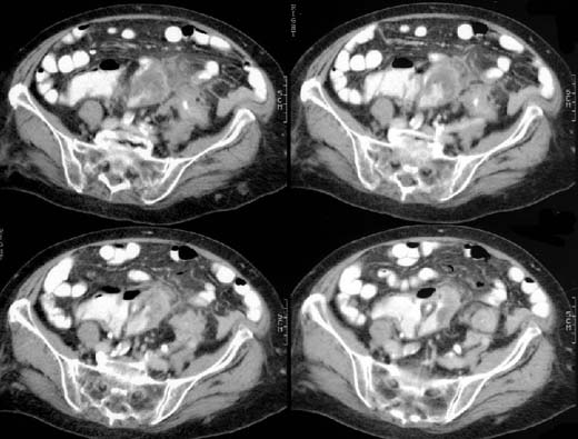
Abdominal CT scan done after i.v., oral and rectal application of contrast
medium.
FINDINGS:
Thickening
of the intestinal wall and narrow
lumen of the sigmoid colon. The adjacent mesenteric
pericolic fatty tissue appears consolidated and contrast-enhancing.
DIAGNOSIS:
Sigmoid
diverticulitis with suspected perforation.
Web
resources:
1) Ultraschallatlas
2) Uni Marburg
3) Chorus
4) Medicine worldwide
5) AHC-Consilium