CASE
HISTORY / CLINIC:
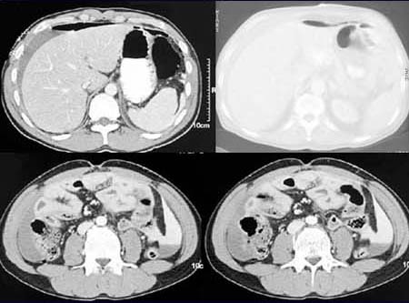
32-year-old patient with severe abdominal pains.
FINDINGS:
Demonstration of free
air below the abdominal wall in the abdominal CT scan done after i.v and
oral application
of contrast medium. Fluid collection (possibly blood) mixed
with contrast material. Pure
contrast material and air-fluid
level in between.
DIAGNOSIS:
Perforated hollow organ, in this case duodenal ulcer, with leakage
of contrast material into the abdominal cavity. Pneumoperitoneum.
Web resources:
1) Medicine worldwide
2) TU München
3) Chorus
4) AHC-Consilium