CASE
HISTORY / CLINIC:
57-year-old patient with hematuria
FINDINGS:
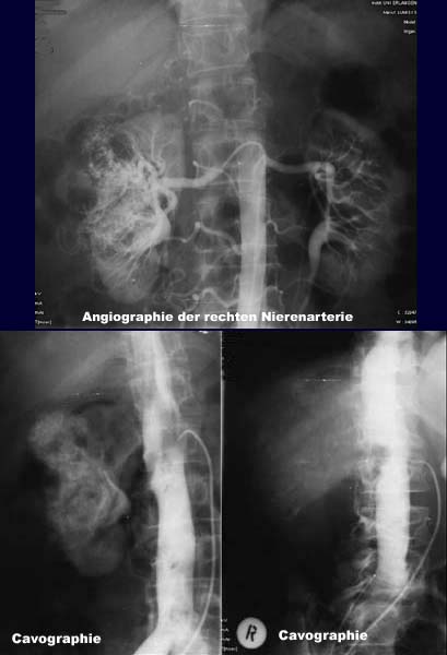
The renal angiography shows evidence of a big hypervasculated
tumor and numerous pathological tumor vessels with arteriovenous fistulas and
invasion of the renal pelvis. The cavography shows a filling
defect in the renal vein and in the inferior vena cava.
DIAGNOSIS:
Big renal
cell carcinoma with tumor
cones in the renal vein and inferior vena cava.
Web resources:
1) AWMF
2) Department of Radiology
3) Chorus
4) Lebertransplantation.de
5) Medicine worldwide
6) AHC-Consilium